


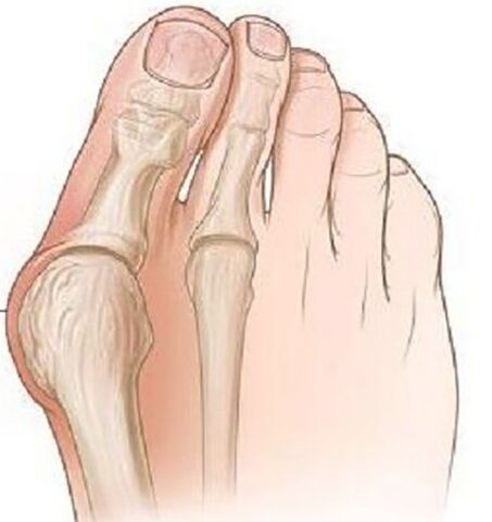
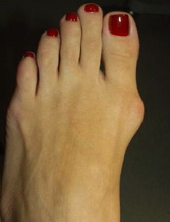
Valgus deformation of the leg thumb is the most common deformation of the foot characterized by the outward difference of the thumb.
Pathology is bilateral in nature, primarily women under the age of 35.In most cases, the deformation is accompanied by chronic inflammation of the joint bag and deformary arthrosis of the plusnephalang joints and the first metatarsal bone pronation differences.
Causes of the disease.Why is it dangerous?
The etiological causes of the thumb of the thumb of the thumb have not yet been fully investigated.There are many occurrences theories:
- Vestigial theory.In the mid -19th century, it was believed that only models were submitted to the models because of the high level of model shoes, but during the study this pathology was found in men who had flat shoes.
- The theory of primary muscle weakness - was refuted in detailing the problem.
- The theory of the weakness of the league and the lack of aponeurosis of the sole - most scientists follow this theory.
There are many factors leading to this pathology debut:
- Excessive weight increases the load on the legs.
- Age -related dyspressive changes in the league joint device.
- For the heel shoes, more than 5 cm, narrowed to the toe.
- Existing deformation of the skeleton (skoliosis, femur and knee joints, flat legs).
The risk of pathology lies in the course of time that this deformation leads to the deformation of posture, may develop a distrofical depth of the spine, hip and knee joints due to improper distribution of the filling during walking, and injury to the foot muscles with constant pain, which significantly destroys the person's well.
Clinical manifestations
At the beginning of the disease, the deformation of the valgus does not show clinically.Most women are more concerned about the appearance of the Metacarpal-Phalanx joint and the outbreak of the first finger becomes noticeable.
Over time, pain occurs, first when wearing narrow shoes and longer walks, and later the pain is constant, painful.
Due to the inadequate distribution of the weight, the limb is formed with the first and second fingers of the sole, the second finger is raised, strongly extended, and the fingers of the "malleus" are greatly complicated.In the anterior part of the blood circulation and the anterior part of the leg, arthrosis and chronic bursitis develop.
Diagnosis
- Objective examination shows the classic deformation with the valgus differences between the thumb, and the distal part of the foot was expanded.The first metatarsal bone head is projected by bursite - hyperemia and skin edema, pain during palpation.
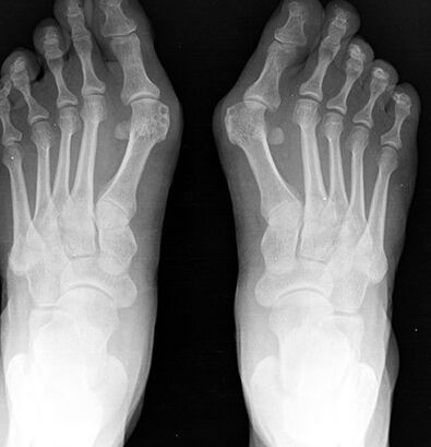
- Radiography of the foot in two projections.The anterior projection determines the degree of thumb, the condition of the metacarpal-phalanx joint, the degree of sasamoid bone displacement, and in the lateral projection, the degree of flat legs is often displayed, which often occurs with the valgus differences of the front fingers of the foot.
- Plantography.When printing the foot on the paper, there is a line in the middle of the heel, between the fourth and the third fingers, the outer set of the foot, based on the presence of the foot and its degree.In these patients, the flat foot or flat foot of 1 degree is more common.
Treatment and prevention
Depending on the severity of the process, conservative or surgical treatment is performed.
Conservative treatment is performed at a slight stage of the disease, various orthopedic insoles are used separately -separately and the deformed finger is placed in the normal position while stabilizing and distributing the load on the front of the limb.
In children and senile age, the transverse knitting of the distal limb is used, lay between the first and the second fingers.To reduce pain, hot baths are used with sea salt and soda, and radon baths provide good results.
For the fight against bursitis, local anti -inflammatory gels, compressions with dimexide and lidocaine are used and used with pronounced pain, novocaine blockade and intraarticular injections of glucocorticoids.
Surgery treatment is possible at any stage of the disease and is performed during local anesthesia, which reduces a list of contraindications to surgery.
In Section 1, bone-cold growth is removed at the inner edge of the bone, while only reducing the pain of the process, but not preventing deformation.
With severe deviation of the finger, flat legs are presented with osteotomy at the bottom of the extra bone of the first finger, with a bone blade to reject the finger to the normal position, with the help of a lavsan tape, the transverse tape of the sole develops.After surgery, the leg is tightly fixed with a bandage for 4 weeks, and during the year the use of individual orthopedic insoles is recommended.
Prevention of the disease is to use comfortable shoes without high heels, the shoes should sit freely on the leg, the tight toe should not cause discomfort in the thumb.If there are deformations of the skeleton, it is advisable to perform preventive tests for the orthopedic in order to identify and slow down the progress of the leg thumb.
Consequences and complications
The complication of the process is the development of Dechländer or the "March" of the foot, which is characterized by acute pain due to microcraction in the combination of foot and lendaginitis.
Due to the constant inflammatory process and mechanical damage, there is a risk of developing malignant tumors of the bone.